Stage 3 Cancer
All about the Stages of Cancer
If you've been told you have stage 3 cancer, you may be very worried, wondering what that means. What exactly DOES stage 3 mean?
It's a way for doctors to "put a label" on how advanced the cancer may be. When a diagnosis of cancer has been made, the next step is is to figure out what stage the tumor is at. This is very important, because it helps to indicate what sort of treatment you should get.
There are several different ways of grading the stages of cancer. The simplest system grades the development of a tumor in 4 levels, starting at 1.
 Your doctor can explain
While this system is simple, (there are only 4 possibilities), it can be TOO simple. On a scale of 1 to 4, stage 3 cancer looks pretty serious.
And it is. BUT the development and spread of a tumor is much more complicated than a number from 1 to 4.
When other factors from alternative grading systems are used, stage 3 cancer can have a number of degrees of "seriousness" all to itself. Some stage 3 cancer tumors will be less serious than others.
It's possible that your stage 3 cancer is less invasive than, say, an aggressive stage 2 cancer.
What tests are needed to decide what stage a cancer is at?
Stage 3 Cancer
Here are some of the tests that may be used to check the stages of cancer:
Chest X-ray.
You may have a chest X-ray to check whether cancer cells have spread to the lungs. This is very unlikely unless your cancer is in the advanced stages. You will also have a chest X-ray if you are going to have surgery under general anaesthetic.
CT scan.
A CT scan is a computerised series of X-rays giving a 3 dimensional picture of an area of the body. You may have a CT scan of your head and neck, which can show the size of the tumor and any enlarged lymph nodes in the neck.
MRI scan.
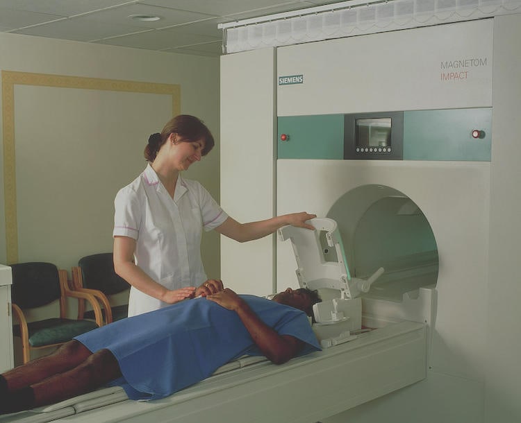
You may have an MRI scan. This stands for Magnetic Resonance Imaging, and it shows soft tissues more clearly than a CT scan. It's very useful for mouth cancers, and can show the degree of spread of a stage 3 cancer.
 Preparing an MRI scan
Ultrasound scan.
An ultrasound scan can look at the lymph nodes in your neck. This scan uses sound waves to create pictures of your body. Again, because stage 3 cancer involves spread of cancer cells to the lymph nodes, an ultrasound scan can be very important in making a diagnosis.
 Ultrasound scan
Panoramic Radiograph.
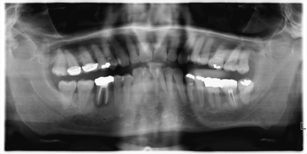
This type of X-ray takes pictures of the area around the upper and lower jaws. It can pick up any signs of cancer in and around these bones. This is also called a Panex, an OPG or OPT.
 Panoramic radiograph
PET-CT scan.
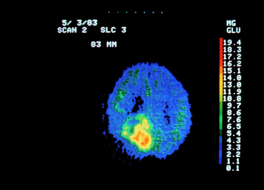
This type of scan is a combination of a CT scan and a PET scan, rolled into one. A PET scan involves injecting a small amount of a radioactive substance which shows up various structures of your body. You may have this type of scan if cancer cells have been found in the lymph glands of your neck, but your doctor doesn't know where they have come from. A PET-CT scan can sometimes help to show a cancer that other scans have not been able to find.
 PET scan image of a brain tumor
A Barium Swallow.
This is not a common test, but you may have one if you are having difficulty swallowing. It shows the area around your vocal chords (larynx) and the top of your oesophagus.You swallow a barium-containing liquid. This shows up very clearly on an X-ray, and anything abnormal will show up.
After your tests, it's normal for you to worry a bit while you are waiting for the results. It's a good idea to try to talk with some of your close family or close friends. It's usually a great help, mentally, if you can share your concerns with someone.
Your test results may take a little time, sometimes a week or two. When they are confirmed, you will need to see your doctor again to discuss what shows up. You and your specialist can then decide on the best course of treatment.
The following stages are frequently used to describe cancer of the mouth, according to the organisation www.oralcancerfoundation.org
Stage 1 cancer.
The cancer is less than 2 centimeters in size (about 1 inch across), and has not spread to lymph nodes in the area. This is very localized and usually easily removed surgically.
Stage 2 cancer.
The cancer is more than 2 centimeters in size, but less than 4 centimeters (less than 2 inches across), and has not spread to lymph nodes in the area. Obviously, a bit more advanced that stage 1, but still frequently treated by surgery and maybe backed up with some radiotherapy or chemotherapy.
Stage 3 cancer.
Either the cancer is more than 4 centimeters in size, OR the cancer is any size but has spread to only one lymph node, on the same side of the neck as the cancer. The lymph node that contains cancer measures no more than 3 centimeters. The important step in classifying a stage 3 cancer is that it has spread to the lymph nodes in the neck.
Stage 4 cancer.
The cancer has spread to tissues around the lip and oral cavity, and the lymph nodes in the area may or may not contain cancer. OR the cancer is any size and has spread to more than one lymph node on the same side of the neck as the cancer, to lymph nodes on one or both sides of the neck, or to any lymph node that measures more than 6 centimeters (over 2 inches). OR the cancer has spread to other parts of the body.
Recurrent
The term "recurrent disease" means that cancer has returned or started up again after previous treatment. It could come back in the lip and oral cavity or in any other part of the body
The TNM system
This is another way of grading the stages of cancer in a more accurate way, but it's more complicated. In this system, stage 3 cancer is basically the same, but the TNM system gives more information about the degree of the spread of the cancer cells.The following information is from www.oralcancerfoundation.org/
T stands for the tumor
N stands for the lymph nodes
M stands for metastasis, where cancer cells have travelled to another part of the body.
The stages of cancer according to this system are as follows:
TX = The primary tumor can't be accurately assessed. This means it's not possible to check it properly. There might be a tumor, there may not be. It's impossible to say either way.
T0 = No evidence of a primary tumor. We can inspect the suspicious area, and make a decision. No tumor.
T i s = Carcinoma in situ. There is a tumor, very small, and it hasn't spread out at all yet.
T1 = A tumor 2 cm or less across. A relatively small tumor.
T2 = A tumor more than 2 cm across, but less than 4 cm.
T3 = A tumor more than 4 cm across, and is showing signs of invading neighboring tissues, such as muscle or bone. This is basically the same as stage 3 cancer.
T4= A tumor that has spread into next-door tissues, for example through the jawbone, or into deep muscle of the tongue.
Then the stages of cancer for the lymph nodes are graded, giving a more detailed (and therefore more accurate) picture of the actual stage that the tumor is at;
N0 = No regional lymph node metastasis. This means that the tumor cells have not spread to any lymph nodes.
N1 = Spread of cancer cells (called metastasis) into a single lymph node, on the same side as the tumor, and it is less than 3 centimeters in greatest dimension
N2 = Metastasis in a single lymph node, on the same side, but more than 3 cm and less than 6 cm in greatest dimension.This is then sub-divided further:
N2a = Metastasis in a single lymph node, more than 3 cm but less than 6 cm across.
N2b = Metastasis in multiple lymph nodes, none more than 6 cm across.
N2c = Metastasis in lymph nodes on the other side of your body than the tumor, but none of them are more than 6 cm in size.
N3 = Metastasis in any lymph node more than 6 cm in size.Next, we have to assess if there are any cancer cells a significant distance away from the main tumor, for example in the lungs or the liver. This is graded with the initial "M".
MX = Metastasis to other organs at a distance from the tumor cannot be assessed, for whatever reason.
M0 = No distant metastasis. There is no evidence of any cancer cells having travelled elsewhere in your body.
M1 = Distant metastasis. There IS evidence of cancer cells at a distant organ from the original tumor.
This grading system gives doctors a quick and reasonably accurate way of describing the stages of any cancer. Given a grading like this, a doctor will be able to assess how much the cancer has spread within seconds.BUT there is one aspect of cancer that these grading systems do not take into account, and that aspect is how aggressive the cancer cells are.
Grade.
This is a way of grading how aggressive a cancer is.
The definitions of the "G" categories apply to all head and neck cancers except thyroid cancer. The term "differentiated" tumor cells means cells that are microscopically similar to normal cells. They tend to grow and spread at a slower rate than undifferentiated or poorly differentiated tumor cells, which don't have the structure and function of normal cells, and grow very rapidly.
Poorly differentiated tumors are able to get across and invade all types of tissue. They can grow through all soft tissues, muscle, and even into bone.
G = Grading according to how the cancer cells look under a microscope, and how they behave.
GX = "Grade of differentiation cannot be assessed". We cannot tell how differentiated the cells are.
G1 = "Well differentiated cells". This is a very slow-growing tumor.
G2 = "Moderately differentiated cells". These cells grow more quickly than normal body cells, but are not particularly aggressive.
G3 = "Poorly differentiated cells". This cancer will grow quickly, and tend to spread.
G4 = "Undifferentiated cells". This is the fastest-growing tumor type, and will spread quickly through all body tissues next to the tumor, as well as metastasizing cancer cells to other parts of the body.
While a lot of this looks like grim reading, there are positive aspects to take away. You may have been told that you have a stage 3 cancer. But that's not the whole story. You need to know the TNM score, too. And even though you may have a stage 3 cancer, it may well be a well-differentiated tumor, which means it progresses slowly. Ask your doctor to explain this to you.


